
|
| mhiptv.org |
|
|||||||
 |
|
|
أدوات الموضوع | انواع عرض الموضوع |
 15-04-2012, 09:24 PM
|
#1 |
|
مدير سابق ومؤسس الموقع
 تاريخ التسجيل: Sep 2009
الدولة: Egypt - Alexandria
المشاركات: 11,880
  |
Bones of the skull
عظام الجمجمة 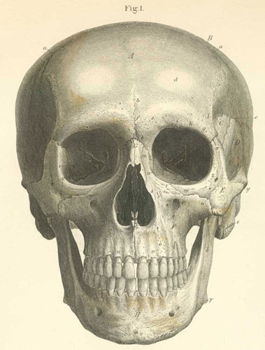 من الامام 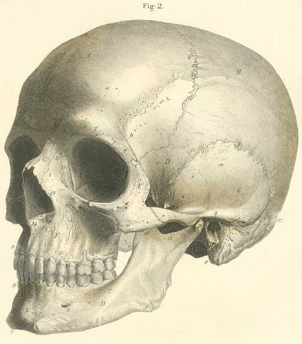 منظر جانبي 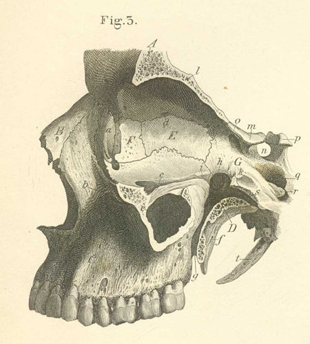 الجدار الداخلي للعظام من المدار، والحفرة الجناحية والمناطق المحيطة بها The inner wall of the bones of the orbit, and the pterygopalatine fossa and their surroundings A. Frontal bone. B. Nasal bone C. Maxillary bone. D. Palatine bone (perpendicular part; pterygopalatine fossa). E. Ethmoidal bone (lamina papyracea). F. Lacrymal bone. G. Sphenoid bone. a) Fossa for lacrymal sac. b) Infraorbital foramen. c) Ethmoidal foramen. e) Maxillary sinus (antrum of Highmore). f) Pterygoid process. g) Pterygopalatine canal. h) Pterygopalatine foramen. i) Orbital process, palatine bone. k) Sphenoid process, palatine bone. l) Orbital part of the frontal bone. m) Anterior clinoid process. n) Sella turcica. o) Optic foramen. p) Posterior clinoid process. q) Carotid canal. r) Lingula. s) Pterygoid canal (Vidian canal). t) Styloid process. 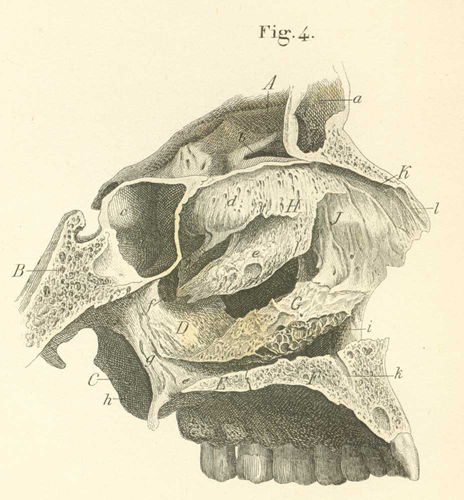 The external wall of the (left) nasal bones with their muscles الجدار الخارجي للعظام الأنف (يسار) مع عضلاتهم ) ) Frontal bone, frontal part. B) Sphenoid bone, body. C) Pterygoid process of the sphenoid bone. D) Palatine bone, perpendicular part. E) Palatine bone, horizontal part. F) Maxillary bone, palatine process. G) Inferior concha, inferior turbinate bone. H) Nasal lamina, ethmoid labyrinth. I) Frontal process (nasalis of the maxillary bone). K) Nasal bone. a) Frontal sinus. b) Ethmoidal sinus. c) Sphenoidal sinus. d) Superior concha, highest turbinate bone. e) Middle concha, middle turbinate bone. f) Pterygopalatine foramen, in the perpendicular part of the palatine bone. g) Pterygoid process, medial wing. h) Pterygoid process, lateral wing. i) Lacrymal canal, exit. k) Incisive canal (for the passage of the nasopalatine artey, vein, and nerve). l) Sulcus for the external nasal branch of the anterior ethmoid nerve 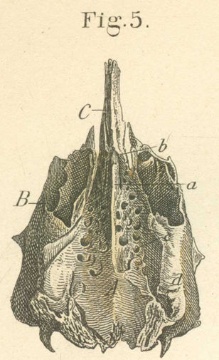 The ethmoid bone, by its upper outer surface العظم الغربالي سطحه الخارجي العلوي  The left palatine bone, from its media surface عظام الحنك من اليسار 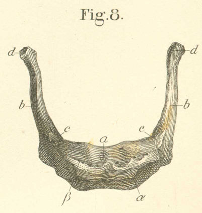 The hyoid bone, from the front. الجبهة- العظام اللامي 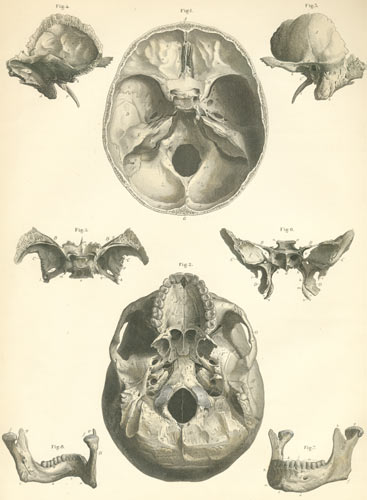 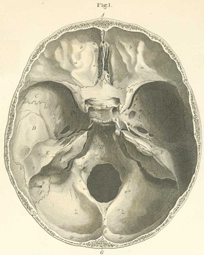 The interior base of the skull القاعدة الداخلية للجمجمة 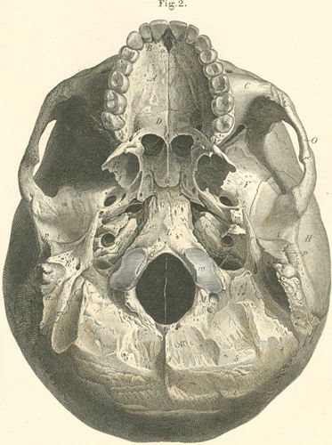 The inferior or outer surface of the skull base السطح السفليالخارجي لقاعدة الجمجمة 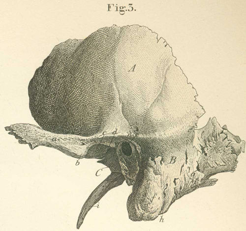 The left temporal bone seen from the outside العظم الصدغي من الخارج 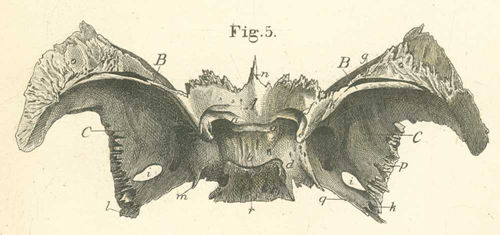 The os sphenoid, from its upper or its brain surface A) Body or base. B) Lesser wing or ensiform process of the sphenoid. C) Greater wing (cerebral outer surface). a) Medial clinoid process. b) Sella Turcica, the fossa for the hypophysis gland. c) Posterior clinoid process. d) Carotid sulcus. e) Anterior clinoid process. f) Optic foramen. g) Superior orbital fissure. h) Foramen rotundum. i) Foramen ovale. k) Foramen spinosum. l) Spinous process. m) Lingula. n) Sphenoid crest. o) Superior margin (articulates with the frontal bone). p) Posterior margin (articulates with squamous part of temporal bone). q) Posterior inferior part (placed on the petrus part of the temporal bone). r) Clivus. 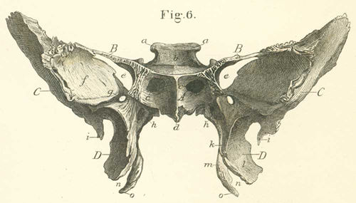 The anterior surface of the sphenoid bone. A) Body of the sphenoid bone. B) Lesser wing of the sphenoid. C) Greater wing of the sphenoid (orbital surface). D) Pterygoid process. a) Posterior clinoid process. b) Sella turcica (fossa for the hypophysis). c) Orifice of the sphenoid sinus. d) Sphenoid crest. e) Superior orbital fissure. f) Orbital surface of the greater wing of the sphenoid bone. g) Foramen rotundum. h) Vidian canal (pterygoid canal). i) Spinous process. k) Pterygoid sulcus or palatine sulcus (the posterior wall of the pterygopalatine canal). l) Lateral wing of the pterygoid process. m) Medial wing of the pterygoid process. n) Pterygoid notch. o) Pterygoid hamulus. 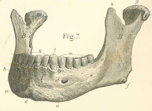 The mandible seen from its anterior surface الفك السفلي السطح الامامي A) Body, horizontal part. B) Ramus. a) Inferior surface of the body; the base (attachment site for mm scalene, quadratus menti, platysma, and digastricus). b) Alveolar margin, with sixteen alveoli. c) External mental protuberance. d) Mental foramen (passage for mental artery, vein, and nerve; exit for the inferior alveolar canal). e) External oblique line (origin for the m buccinator). f) Mandibular angle (insertion for m masseter). g) Condyloid process (from the neck). h) Coronoid process (insertion for m temporalis). i) Semilunar incisure. k) Incisure teeth. l) Canine teeth. m) Molar teeth. 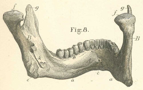 The mandible is placed to see the inner surface السطح الداخلي للفك السفلي A) Body or horizontal part of the mandible. B) Ramus of the mandible. a) Base or inferior surface. b) Alveolar margin. c) Internal mental spine (origin for mm geniohyoid and genioglossus). d) Internal oblique line (origin for mm mylohyoid and for mylopharyngeus or superior pharyngeal constrictor). e) Inferior mandibular angle. f) Condyloid process. g) Coronoid process. h) Semilunar incisure. i) Inferior alveolar foramen (entrance to inferior alveolar canal). k) Mylohyoid sulcus, for the mylohyoid nerve from the inferior alveolar nerve Bones of the trunk 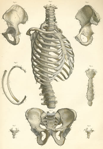 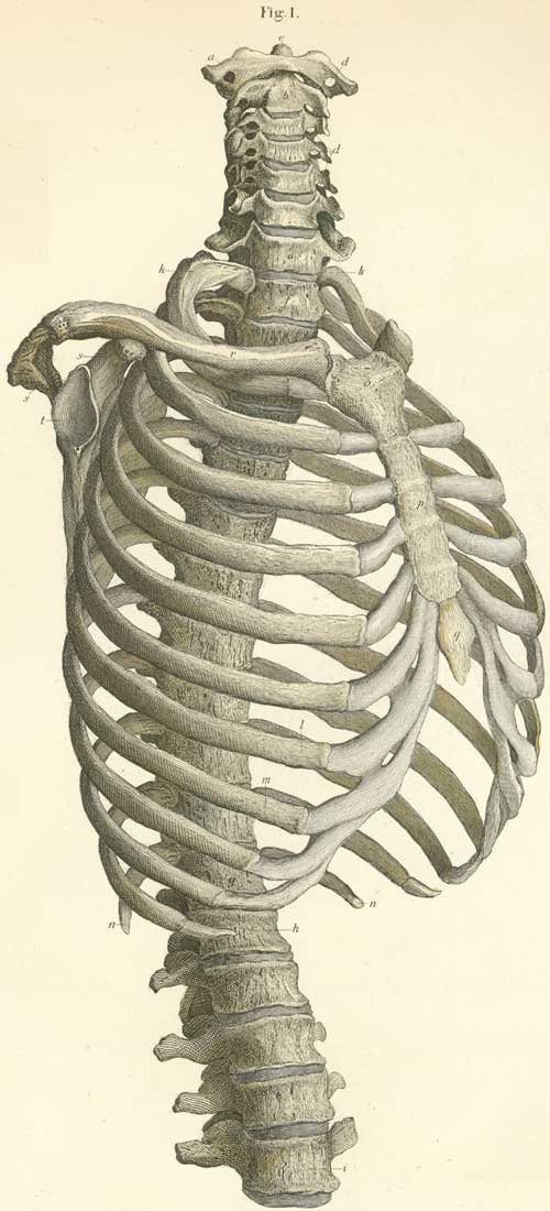 The vertebrae of the thorax, with the cervical, lumbar and shoulder; on the right side and seen from the front a) Atlas, first cervical vertebra. b) Axis (epistropheus), second cervical vertebra. c) Seventh cervical vertebra. d) Vertebral canal. e) Odontoid process of the axis. f) First thoracic vertebra. g) Twelfth thoracic vertebra. h) First lumbar vertebra. i) Fifth lumbar vertebra. k) First (true) rib. l) Seventh (true) rib. m) First false or eighth rib. n) Last (twelfth) rib. o) Manubrium sterni. p) Body of sternum. q) Xiphoid process sterni. r) Clavicle. s) Scapula. t) Glenoid cavity of scapula (for the head of the humerus 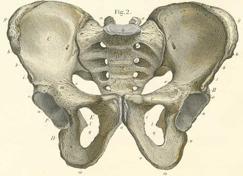 The bones of the pelvis, from its anterior side عظام الحوض - جانبي A) Sacral bone (os). B) Coxal bone. C) Os ilium. D) Os ischii. E) Os pubis. a) Superior oblique process of sacral bone. b) Body of the sacral bone (with the prominence [sacral vertebral angle]). c) Internal arcuate line (marks true from false pelvis). d) Anterior sacral foramen. e) Internal arcuate line of the ilium. f) Sacro-iliac symphysis. g) Iliac crest. h) Anterior superior iliac spine. i) Anterior inferior iliac spine. k) Anterior semilunar incisure of the iliac bone. l) Ischial spine. m) Iliopectinate tubercle (pubic tubercle). n) Acetabulum. o) Acetabular lip p) Acetabular notch. q) Obturator foramen. r) Horizontal ramus of the pelvic bone. s) Pubic spine. t) Descending ramus of the pubic bone. u) Symphysis of the pubic bone (beneath, the pubic angle or arch). v) Ascending ramus of the ischial bone. w) Ischial tuberosity. x) Descending ramus of the ischial bone 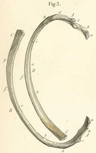 Two ribs seen from the lateral and medial sides A) Posterior extremity of rib. B) Body of the rib. C) Anterior extremity of rib. a) Capitulum (head). b) Collum (Neck). c) Tubercle. d) Angle. e) Upper margin. f) Lower margin. g) Outer surface. h) Inner surface. i) Costal sulcus or groove for intercostal artery, vein, and nerve 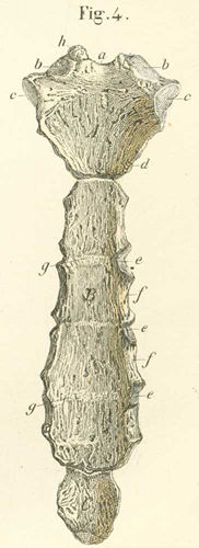 The sternum seen from its anterior surface A) Manubrium. B) Body. C) Xiphoid (ensiform) process. a) Superior semilunar incisure (sternal notch). b) Clavicular cavity or notch. c) Notch for 1st rib. d) Notch for 2nd rib. e) Costal notches (articular fovea). f) Lateral semilunar notches. g) Transverse line. h) Suprasternal ossiculum. 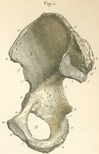 The right innominate (coxal) bone, seen from within, and through the lines of union of the ilium, ischium, and pubic bones A. Ilium. B. Ischium. C. Pubis. D. Obturator foramen (passage by a small aperature for the obturator artery, vein, and nerve). a) Iliac crest (origin for mm quadratus femoris and latissimous dorsi; with an external lip for m external abdominal oblique, internal lip for m. internal oblique and intermediate line for m transverses abdominus). b) Anterior superior iliac spine (origin for mm sartorius and tensor fasciae latae). c) Anterior inferior iliac spine (origin for m rectus femoris). d) Posterior superior iliac spine. e) Posterior inferior iliac spine. f) Anterior semilunar notch. g) Posterior semilunar notch. h) Tubercle of the iliac bone. i) Auricular face (formed with the auricular face of the sacral bone of the sacral ilica symphysis). k) Internal arcuate or terminal line (or the line demarking the greater and lesser (or true) pelvis). l) Greater sciatic notch (passage for m piriformis and inferior gluteal artery, vein and nn inferior gluteal, sciatic, and pudendal). m) Body of the ischial bone (with the continuation of the greater sciatic notch). n) Descending ramus of the ischium. o) Ascending ramus of the ischium (origin for mm ischiocavernosus, gracilis, adductor magnus, and transversus prostatae). p) Ischial spine (origin for sacrospinous ligament, mm gemulus superior and coccygeus). q) Ischial tuberosity (origin for sacrotuberous ligament, mm gemulus inferior, quadatus femoris, biceps femoris, semitendinosus, semimembranosus, adductor magnus, tranverse pernei profundus). r) Lesser sciatic foramen (passage for m obturator internus, artery, vein and internal pudendal nerve). s) Horizontal ramus of the pubic bone. t) Descending ramus of the pubic bone. u) Iliopectineal tuberosity. v) Pubic crest (origin of m pectineus). w) Pubic spine (attachment for the inguinal ligament). x) Pubic symphysis. y) Iliac fossa (inner concave surface of the ilium and the origin of m. internal iliac 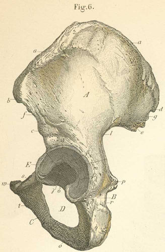 The left pelvis, seen from the outside A) Ilium. B) Ischium. C) Pubis. D) Obturator foramen (passage by a small aperature for the obturator artery, vein, and nerve). E) Acetabulum (for the head of the femoral bone). a) Iliac crest (origin for mm quadratus femoris and latissimous dorsi; with an external lip for m external abdominal oblique, internal lip for m. internal oblique and intermediate line for m transverses abdominus). b) Anterior superior iliac spine (origin for mm sartorius and tensor fasciae latae). c) Anterior inferior iliac spine (origin for m rectus femoris). d) Posterior superior iliac spine. e) Posterior inferior iliac spine. f) Anterior semilunar notch. g) Posterior semilunar notch. h) Tubercle of the iliac bone. i) Auricular face (formed with the auricular face of the sacral bone of the sacral ilica symphysis). k) Internal arcuate or terminal line (or the line demarking the greater and lesser (or true) pelvis). l) Greater sciatic notch (passage for m piriformis and inferior gluteal artery, vein and nn inferior gluteal, sciatic, and pudendal). m) Body of the ischial bone (with the continuation of the greater sciatic notch). n) Descending ramus of the ischium. o) Ascending ramus of the ischium (origin for mm ischiocavernosus, gracilis, adductor magnus, and transversus prostatae). p) Ischial spine (origin for sacrospinous ligament, mm gemulus superior and coccygeus). q) Ischial tuberosity (origin for sacrotuberous ligament, mm gemulus inferior, quadatus femoris, biceps femoris, semitendinosus, semimembranosus, adductor magnus, tranverse pernei profundus). r) Lesser sciatic foramen (passage for m obturator internus, artery, vein and internal pudendal nerve). s) Horizontal ramus of the pubic bone. t) Descending ramus of the pubic bone. u) Iliopectineal tuberosity. v) Pubic crest (origin of m pectineus). w) Pubic spine (attachment for the inguinal ligament). x) Pubic symphysis. y) Iliac fossa (inner concave surface of the ilium and the origin of m. internal iliac). a) Acetabular supercilium. b) Lunate face (with two horns). g) Fossa for ligament capitis femoris. d) Acetabular notch 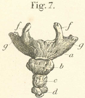 The os coccygis seen from the front a) – d) First to fourth (false) vertebrae. e) Articular cavity ( for the point of the sacral bone). f) Coccygeal cornu. g) Transverse process 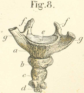 The os coccygis seen from behind a) – d) First to fourth (false) vertebrae. e) Articular cavity ( for the point of the sacral bone). f) Coccygeal cornu. g) Transverse process Bones of the trunk 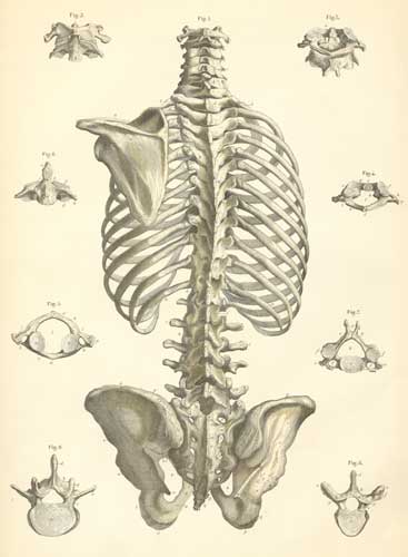 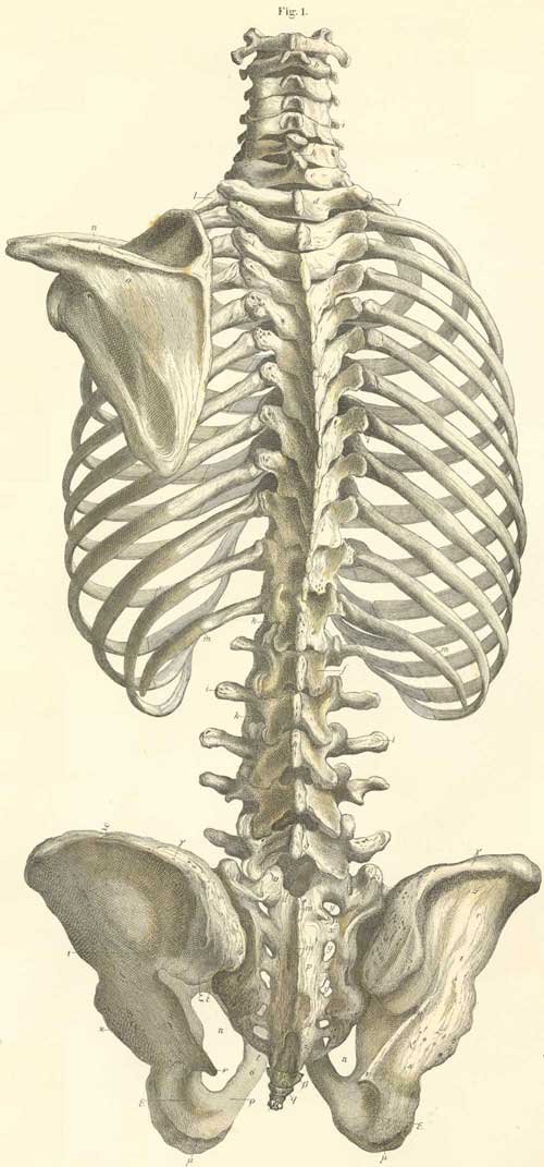 The bones of the trunk with the left scapula, seen from the back a) First cervical vertebra, atlas. (See also figures
b) Second cervical vertebra, axis (epistropheus). (See also figures
c) Seventh cervical (last) vertebra (vertebra prominens). d) First thoracic vertebra. e) Twelfth (last) thoracic vertebra. f) First lumbar vertebra. g) Fifth (last) lumbar vertebra. h) Spinous process. i) Transverse process. k) Intervertebral foramen. l) First (true) rib. m) Twelfth rib. n) Clavicle. o) Scapula. p) Sacral bone. q) Coccygeal bone. r) Ilium (part of the innominate bone). s) Ischium (part of the innominate bone). t) Pubic bone (part of the innominate [coxal] bone). u) Sacral canal (entrance). v) Superior oblique process of the sacral bone. w) False spinous process. x) Spurious oblique and transverse process. y) Posterior sacral foramina. z) Sacral horn. a) Exit of sacral canal. b) coccygeal horn. g) Iliac crest. d) Posterior superior iliac spine. e) Posterior semilunar incisure of the ilium. z) Posterior inferior iliac spine. h) Greater sciatic notch. q) Semicircular line (superior external arcuate line). i)Arcuate line (inferior arcuate line). k) Acetabulum. l) Descending ramus of the arcuate line. m) Ischial tuberosity. x) Lesser sciatic foramen. p) Obturator foramen. r) Ascending ramus of the ischium. s) Descending ramus of the pubic bone (see also Plate 3, figures
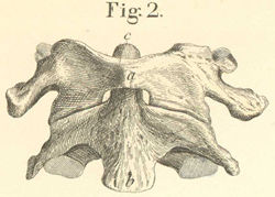 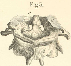 The first two cervical vertebra, Atlas and Axis, seen from the front and back a) Atlas (first cervical vertebra). b) Axis (second cervical vertebra) reversed. c) Odontoid process of the axis. d) Condyloid process of the atlas 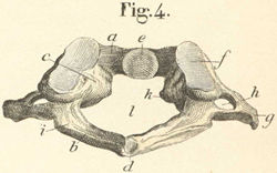 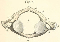 The first cervical vertebra, Atlas, seen from above and behind and from below a) Anterior arch of the atlas (attachment site for rectus capitis anterior minor, anterior atlanto-occipital ligament). b) Posterior arch. c) Lateral mass. d) Posterior tubercle of the atlas (origin of rectus capitis posterior minor). e) Fovea dentis atlantis. f) Condyloid fossa of the superior oblique process (formed by the condyle of the occipital bone; a hinge joint). g) Transverse atlantic process (attachment site for mm rectus capitis lateralis, superior and inferior capitis oblique). h) Vertebral foramen (transverse foramen) (vertebral canal for the vertebral artery. i) Groove for the vertebral artery. k) Lateral tubercle for ligamentum transversum. (lig. Cruciforme atlantis). l) Vertebral foramen or canal. m) Anterior atlantic tubercle (attachment for m. longus colli, and anterior longitudinal ligament 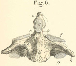 The second cervical vertebra, axis, seen from the front a) Body of the axis. b) Odontoin process. c) Neck of the odontoid process. d) Fovea for dentis atlantis. e) Apex of the odontoid process. f) Superior oblique process (it forms, with the atlas, a rotation joint). g) Inferior oblique process. h) Transverse process, with the vertebral foramen or vertebral canal for the vertebral canal 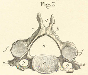 This figure is of a cervical vertebra seen from the front a) Body. b) Arch. c) Spinous process (short and broad). d) Notch in the spinous process for ligamentum nuchae. e) Transverse process (broad, with a groove and hole). f) Superior oblique process (above the continuation of the joint). g) Vertebral foramen (the 7 vertebral foramina for the vertebral arteries and veins. h) Spinal foramen and canal. 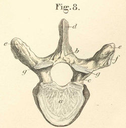 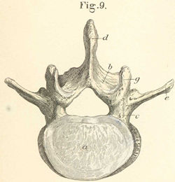 A thoracic (fig. 8) and a lumbar (fig. 9) vertebra. a) Body. b) Arch. c) Vertebral notch (forms an intervertebral foramen for spinal nerves and branches of spinal arteries). d) Spinous process. e) Transverse process. f) Joint surface for a rib tubercle. g) Superior oblique process. المصدر: mhiptv.org/forums hgid;g hgu/ln hgjavdpn g[sl hgHkshk
__________________
   |

|

|
|
16-04-2012, 02:54 AM
|
#2 |
|
مشرف سابق
تاريخ التسجيل: Aug 2011
المشاركات: 2,566
|
جزاك الله الف خير على كل ما تقدمه
|
|
|
|
|
24-09-2012, 10:09 AM
|
#3 |
|
:: عضو محترف ::
تاريخ التسجيل: Sep 2012
المشاركات: 262
|
مشكووووووووووووووووور
|
|
|
|
|
|
|
 المواضيع المتشابهه
|
||||
| الموضوع | كاتب الموضوع | المنتدى | مشاركات | آخر مشاركة |
| الهيكل العظمي البشري | جولد 2020 | القسم الطبي العام | 0 | 16-06-2011 01:17 AM |
| محام بالبحيرة يقاضى فتحي سرور ويتهمه بالخيانة العظمى | soliman2 | قسم الأخبار والصحافة العالمية | 0 | 03-03-2011 09:07 PM |
| الدول العظمى العشرين خيبت الآمال 25/10/2010 caya | traderxp | قسم الإقتصاد والمال | 0 | 25-10-2010 09:56 PM |
| القصة الكامله .. لاقامة الهيكل المزعوم .. وهدم الأقصى بأيدى اليهود | soliman2 | قسم الأخبار والصحافة العالمية | 0 | 20-03-2010 10:28 PM |
| التركيب التشريحى للقدم والكاحل | soliman2 | قسم المعلومات و النصائح الطبية | 1 | 08-03-2010 02:36 AM |


 العرض العادي

